Contents |
Heidelberg Retina Tomograph: Better Information, Better Control of Glaucoma
Introduction
Glaucoma is an eye disease most commonly characterized by increased pressure within the eye, known as intraocular pressure (IOP). While increased pressure is one important clinical sign, glaucoma is more complex than a simple measure of pressure.
Instead, it is helpful to consider glaucoma as a disease affecting the optic nerve and all the individual retinal nerve fibers that comprise it.
In the early stages, glaucoma has no symptoms and often goes undiagnosed and untreated because of that lack of symptoms. It is understandable that if one does not know of the existence of a medical problem, no examination, diagnosis or treatment is sought.
Until recently, testing for glaucoma included measuring IOP and evaluating vision in the peripheral areas of vision with testing of visual fields. Unfortunately, documenting progressive vision loss is only possible once loss has begun to occur; even more unfortunately, vision loss from glaucoma is not reversible.
Heidelberg Retinal Tomograph
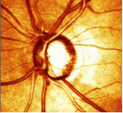
Recent developments in computer imaging technology now allow for the sensitive nerve fiber layer of the retina and the optic nerve to be evaluated using a three-dimensional cross-section view. The Heidelberg Retinal Tomograph (HRT) is a system combining a laser-scanning camera and specialized software that allows evaluation of the optic nerve itself.
This revolutionary technology allows for much more understanding of the progression of optic nerve, not only in glaucoma but other eye conditions as well, and it allows this long before irreversible vision loss occurs. (See figure.1)
Tomography uses real-time information gathering from the living eye for immediate study and analysis. In addition, each captured image is compared with a database of normal outcomes for each patient’s age. It has been shown that 3-D measurements of the optic nerve head are far better than conventional examination methods such as visual fields testing and IOP alone.
How does it Work?
The HRT works something like an ultrasonic scanner, except instead of using sound, the process uses reflected light and converts this into a layered image with enhanced colors indicating various thicknesses of tissue.
Dilation of the pupil is not usually needed, and the HRT exam takes only a few minutes; it is painless and non-invasive.
This technology allows for a much more accurate assessment of disease, its progression and treatment options. It has now become the standard of care for patients with documented glaucoma, as well as those with other risk factors, which include:
- High myopia (nearsightedness)
- High hyperopia (farsightedness)
- Shallow anterior chamber on examination
- Regular long-term steroid use
- Previous eye injury
- Age over 45 years
- Any family history of any type of glaucoma
- Diabetes
- African descent
Glaucoma is one of the leading causes of blindness throughout Canada and the US. The best way to control it and prevent vision loss is to catch it early. People should be aware of their risk factors and be sure to have a thorough eye examination at appropriate intervals as recommended by an eyecare practitioner.
Glaucoma is painless, but so is testing and treatment if needed. No one should ever lose vision to untreated glaucoma.

